Captured in the Endosome
In medicine, biologically active macromolecules as therapeutics, such as mRNAs, are becoming a widely used strategy. Cells take up macromolecules via the endocytic pathway and traffic them to their specific site of action within the cell. Currently, uptake into endosomes, endosomal distribution, and escape of molecules from endosomes into the cytoplasm are still poorly understood and challenging steps in drug development and delivery. Therefore, we have applied our know-how of endosomal transport to explore the uptake mechanisms and endosomal escape in collaboration with pharmaceutical companies. Our expertise in endosomal trafficking and membrane fusion allows us to develop novel macro molecule delivery systems based on bio-inspired approaches.

The cell takes up delivery vehicles, such as lipid nanoparticles (LNPs), via the endocytic pathway. The active compound packaged within the LNP is released from the LNP and escapes from inside the endosome into the cytosol.
Understanding the Mechanism of Endosomal Escape
In collaboration with Alnylam Pharmaceuticals, we characterize the delivery of siRNAs formulated in lipid nanoparticles (LNPs) to silence genes in cells and mouse liver. LNPs, the vehicle also used for some Covid19 mRNA vaccines, are small particles composed of different lipid types that wrap the RNA molecule and help to cross cell membranes. We used LNPs containing siRNAs connected to small gold particles and followed cellular trafficking using electron microscopy. Our quantification showed that only 1-2 % of all siRNAs escape from the endosomes into the cytosol both in cells and in mouse liver. Furthermore, siRNAs only escape from the endosomes to the cytosol when the LNP-siRNAs stay in a specific endocytic compartment sharing early and late endosomal characteristics (Gilleron et al., 2013). In a follow-up study with Alnylam Pharmaceuticals, we identified small molecules that improve siRNA delivery to cells (Gilleron et al., 2015).
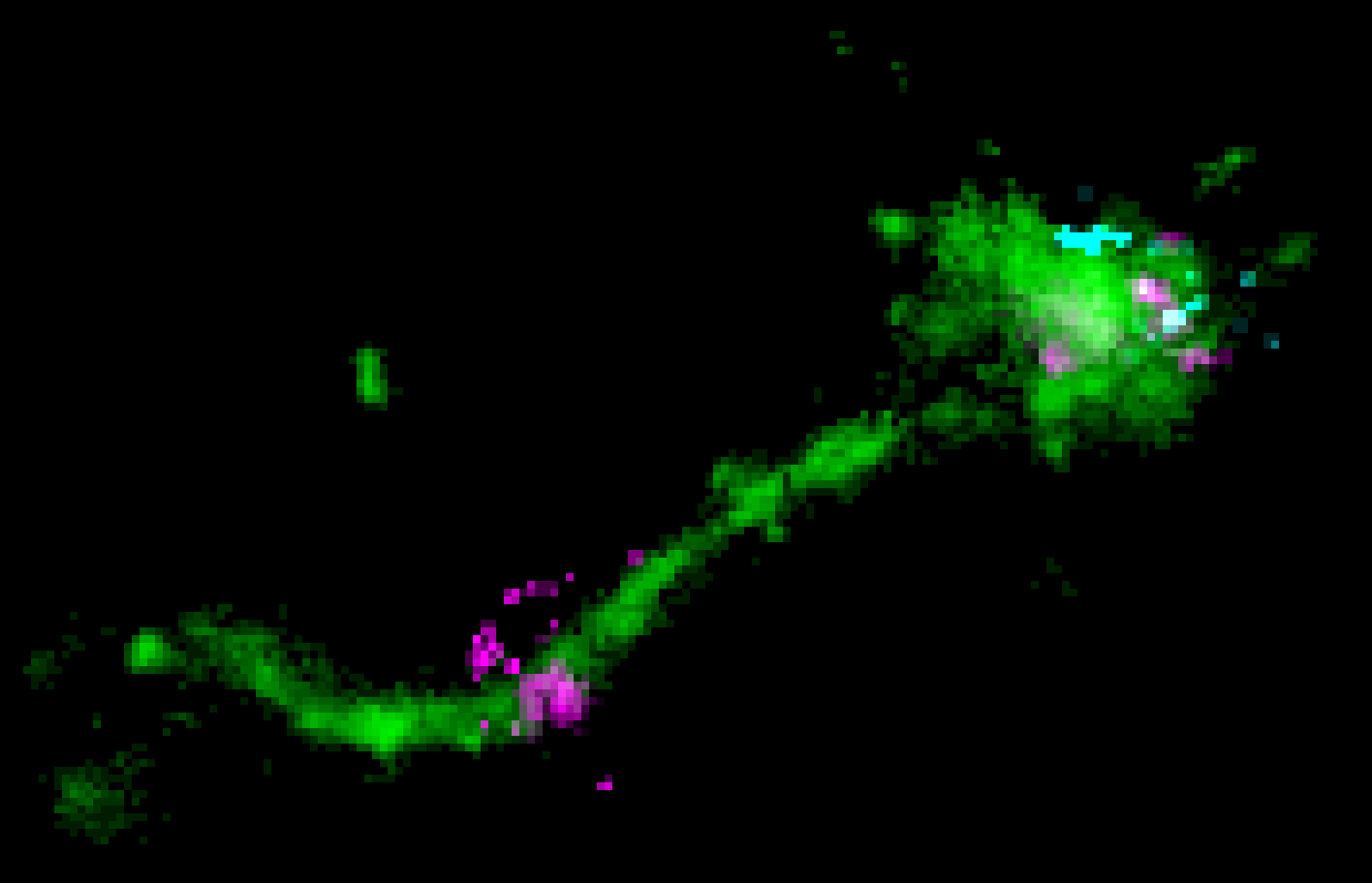
In collaboration with AstraZeneca, we quantified the endosomal escape of mRNA formulated in LNPs in HeLa and primary human cells (Paramasivam et al., 2021; Paramasivam et al., 2022). We used super-resolution microscopy to see single LNP-mRNA particles inside the endosomes and caught escape events of mRNA. This happened especially from endosomal recycling tubules, and not as previously thought from late endosomes. This contradicts the idea in the field that mRNA molecules depend on the acidic environment of late endosomes to escape. Additionally, we found mRNA-LNP particles accumulating over time in large late endosomal compartments impairing their acidification. These compartments were defective in cargo transport and delivery and might even be toxic to the cell (Paramasivam et al., 2022).

An LNP is located on a long endosomal tubule together with a perpendicular dispersed mRNA signal, likely representing an instance of mRNA escape. Modified after Paramasivam et. al., 2022.